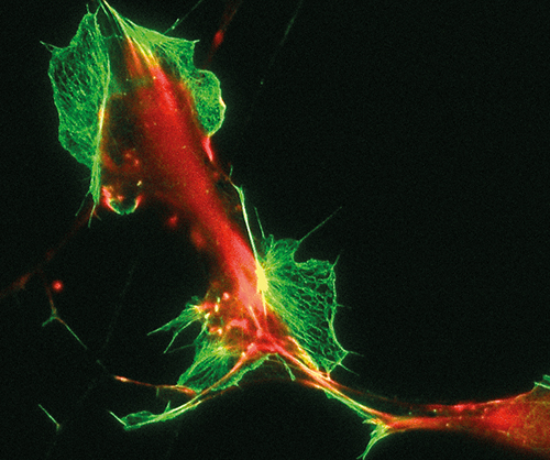
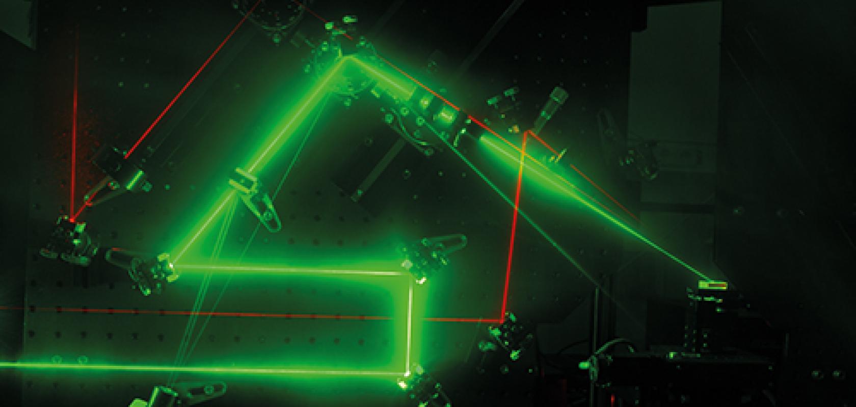
With the ability to produce pulses only picoseconds, femtoseconds, or even attoseconds in duration, ultrafast lasers are able to deliver intense beams of energy to a focal point without causing damage to the materials they are targeting.
This has led to a widespread adoption by researchers in the life sciences field, as it permits the ever more detailed observation of live tissues without the samples being destroyed.
Ultrafast laser manufacturers are now improving these techniques by offering lasers of higher power, longer wavelength and narrower linewidth, which will allow researchers to reach larger depths and levels of detail when examining their live tissue samples.
In the right place at the right time
By being able to deliver high photon densities in their intense beams, ultrafast lasers can be used to perform multi-photon three-dimensional imaging. This technique involves the use of a laser pulse focused through the optics of a microscope to instigate the absorption of two or more photons at the exact same time and location in a live tissue sample, creating an excitation effect that can be observed and interpreted. Combining this with other techniques such as optogenetics, it is possible to excite neurons at certain depths in a brain tissue sample and image the surrounding areas.
‘For example, if a mouse is watching a video, you can image what’s happening in the brain,’ explained Herman Chui, senior director of product marketing at Spectra-Physics. ‘The primary purpose in this case is to study the brain’s structure and function, which could lead to a solution of solving neuro degenerative diseases.’
According to Chui, if a continuous-wave laser were to be used in a standard confocal microscopy setup, fluorescence would come from everything above and below the focal point of the laser, as the excitation wouldn’t be localised, which generates background noise in the imaging process. For a multiphoton process using ultrafast lasers, the only place where the excitation could occur is extremely localised, due to the photons having to converge at an exact location, therefore preventing background noise and enabling clearer imaging.
Ultrafast lasers are the only way to achieve this type of imaging, which provides a very significant opportunity for the devices’ manufacturers. ‘For us it’s one of the four main areas that our ultrafast lasers are used in,’ Chui explained, the others being medicine, micromachining and high-energy physics. Spectra-Physics predominantly provides femtosecond lasers for life science applications, with picosecond lasers more commonly being used for industrial micromachining applications, according to Chui.
While bio-imaging isn’t a new application for ultrafast lasers, new techniques are being developed in the field to increase its capability.

Time-resolved microscopy images of live tissue taken at sub-mllimetre depths

‘There’s a lot of development focused on it,’ confirmed Chui. ‘For the multiphoton techniques, people are moving from using two-photon excitation to three-photon excitation, which requires three photons to be in the exact same place and time to produce an excitation. With that you get even more localised excitations and can image at higher depths into tissue.’
With two-photon excitation, depths of up to approximately 1mm can be imaged in live tissue. However, by switching to three-photon excitation, researchers could potentially reach depths of over a millimetre, which in a mouse brain would enable the imaging of the emotional and memory centres. ‘This is one very interesting direction that has developed in the last year or two,’ Chui commented.
To aid in increasing the depth of multiphoton imaging, manufacturers are now working to offer ultrafast lasers with longer wavelengths. While traditional imaging is performed using green fluorescent protein, which gets excited at around 900nm in a two-photon setup, the light can become scattered at this wavelength as it goes deeper in the tissue. When a longer wavelength is used, however, scattering drops off proportionally and a larger depth of imaging can be reached.
‘There’s a lot of work in moving to longer wavelengths with the two-photon technique. Sources that both we and others have developed are moving out to 1.3μm and are producing 100fs pulses,’ said Chui. ‘With three-photon techniques, people are looking to move further out from 1.3μm to 1.7μm in the future.’
Increasing the power of these longer wavelength lasers is also important, in order to improve their imaging quality. However, manufacturers must do this within the limitations of live tissue, as a significant increase in power could lead to it being damaged when examined. Additionally, because of the large amounts of water in live tissue cells, care must also be taken when choosing higher wavelengths to select those that aren’t significantly absorbed by water, to ensure that imaging can be performed effectively.
Earlier this year Spectra-Physics released its Insight X3 ultrafast laser for multi-photon imaging, a single-source device with a tuning range between 680nm to 1,300nm, nearly double that of Ti:sapphire ultrafast lasers. Suited to delivering higher powers at longer wavelengths – more than 2W at 900nm and 1.4W at 1,200nm – the Insight X3 fits within the limitations of live tissue imaging, and has apparently been well received by the company’s research customers, according to Chui.
Higher power ultrafast lasers can also be coupled with optical parametric amplifiers (OPAs) to deliver suitable wavelengths for multiphoton imaging. Back in June, Spectra-Physics’ new 100W Spirit 1030-100 industrial femtosecond hybrid fibre laser was featured at Laser World of Photonics in Munich. When coupled with the company’s Spirit-OPA and Spirit-NOPA, the system delivers a high-energy tuneable laser output across the visible and infrared spectrum for three-photon deep-tissue bio-imaging and ultrafast spectroscopy applications.
The fastest of flashes
Ultrafast lasers provide numerous advantages over conventional imaging tools in microscopy applications, specifically time-resolved microscopy, which involves taking multiple high-resolution pictures of a sample one after the other in quick succession.
‘A normal camera’s ability to take a series of pictures quickly is limited by the mechanical speed of its rotating shutter, whereas ultrafast lasers feature no moving parts,’ explained Dr Tim Paasch-Colberg, director of marketing for Toptica Photonics, whose ultrafast lasers are often used for time-resolved microscopy. ‘They can therefore achieve much faster imaging rates…as most of them operate at 20-80 million pulses per second, so in principle many images can be taken each second.’ Achieving such speeds enables the study of processes on very fast time scale, for example the chemical processes occurring in live biological samples.

Toptica’s new Femto Fiber Ultra series permits femtosecond pulses with up to 5W output power at 789nm or 1,050nm
In principle there are two main wavelengths used in time-resolved microscopy according to Paasch-Colberg, 780nm and 1,050nm, as between them they can be used to study a broad variety of samples. Toptica Photonics offers both of these wavelengths in its FemtoFiber ultra product line, which was released only recently, at repetition rates of 80MHz and pulse widths of <150fs and <120fs respectively.
‘Many ultrafast microscopy techniques also require a lot of power, and these two machines are at around the highest powers currently available at these wavelengths – up to 500mW for 780nm and up to 5W at 1,050nm,’ Paasch-Colberg said.
According to Paasch-Colberg, a key feature sought after by researchers using ultrafast lasers is the ability to operate them hands-free without difficulty, having them act simply as another tool in their experiment. Manufactures therefore design ultrafast lasers to work completely maintenance-free and operate as a compact box that can be turned on when needed. This small footprint and simple functionality therefore makes them easy to integrate into a wide range of equipment setups.

Toptica’s ultrafast lasers are based on fibre technology
Probing the surface
Lithuanian manufacturer Ekspla considers itself a specialist in picosecond lasers, for which it targets applications involving non-linear phenomena, such as sum frequency generation (SFG) vibrational spectroscopy, a highly sensitive technique used to analyse surfaces. The process uses two beams, one in the visible and one in the mid-IR region, that spatially and temporally overlap at the surface of a material. An output beam is then generated at a frequency equal to the sum of the two input beam frequencies, leaving the surface to be picked up by a detector.
‘We develop special devices for this application,’ said Dr Zenonas Kuprionis, commercial director at Ekspla. ‘A single laser is used to produce both beams. One beam is going directly from the laser, and the same laser is used to pump an optical parametric amplifier. Both beams are precisely synchronised. In this particular application the visible beam is used to convert information coded in the absorption of the IR beam to the spectral range in which photon detectors are the most sensitive
The picosecond laser provided by Ekspla for SFG vibrational spectroscopy is its PL2230 series, a diode-pumped high energy picosecond Nd:YAG laser offering 20-80ps 35mJ pulses at a 50-100Hz repetition rate. The firm also produces its PG501 optical parametric amplifier for use in SFG vibrational spectroscopy, in addition to complete SFG spectrometer systems.
According to Kuprionis, one of the most important laser features for non-linear surface spectroscopy applications is the spectrum linewidth of the tuneable linewidth sources used. Ekspla is therefore trying to make this as narrow as possible in order to distinguish the different vibrational features of live tissue samples.
‘To achieve a narrow spectrum line from OPAs you need to be very careful with the pumping laser line,’ said Kuprionis, who explained that the spectrum of ultrashort pulses broadens because of non-linear interactions, which need to be avoided. ‘You need to apply some special techniques in optical parametric amplifiers to narrow the generation spectra. We use spectral filtration by diffraction gratings or synchronously pumped optical parametric oscillators in this case,’ he said.
While femtosecond lasers do offer high temporal resolution, their spectra are usually too broad for probing molecules, according to Kuprionis, which is why Ekspla is selling more and more tuneable picosecond lasers that are synchronised with femtosecond lasers for this purpose. ‘This allows scientists to achieve more accurate and precise results,’ he commented.

Ekspla’s sum frequency generation microscope provides the ability to investigate spatial and chemical variations across the surface as a function of time
Out of the various emerging advancements in laser design that have occurred, for Ekspla, according to Kuprionis, the most significant is the development of OPCPA (optical parametrical chirped pulse amplification) technology. ‘For us it’s a very important area because our picosecond lasers are very suitable to pump OPCPAs,’ he said. ‘With our partners Light Conversion and National Energetic, we develop ultra-high intensity ultrafast OPCPAs.’
With these partners, Ekspla is producing OPCPAs for the European Extreme Light Infrastructure (ELI) project, which aims to develop and offer the world’s most intense laser systems and make them available to the international scientific community. Ekspla has already produced a five terawatt, 1kHz OPCPA-based laser system worth €4 million with Light Conversion for the project. Now it is developing an additional terawatt laser system with 6fs pulse duration with Light Conversion, and an additional 10-petawatt laser system with National Energetics.
‘There are completely new types of applications for these sources,’ commented Kuprionis. ‘These are extreme light applications like fusion energy experiments, compact high gradient electron and proton accelerators. Also with these sources it’s possible to generate ultrafast x-rays for structural studies of solids and molecules and to create extreme states of matter.’ These extreme sources can also be used for generating pulses only attoseconds in duration, a completely new area of physics that is just at its beginning.
Lasers of the future
While ultrafast lasers are currently around the size of two shoeboxes – one containing the laser, the other the supply electronics – Paasch-Colberg, of Toptica Photonics, believes that they will be shrunk down even further in the coming years.
‘I think the future will lead to size reductions without lowering the performance or changing the important laser parameters (wavelength, pulse duration etc),’ he said. ‘Micro optics will be a key technology for reducing the size of lasers, as well as photonic integrated circuits. Shrinking the components down to a smaller chip while maintaining the same level of power and other specifications will be a big true challenge. I think for biophotonics applications [however], this will be the future.’
Kuprionis, of Ekspla, believes that hybrid laser technology is set to have a positive impact on ultrafast lasers, a laser format already used in micromachining applications and for OPCPA pumping. ‘This involves using a fibre-based seeder, which gives you a lot of opportunities to choose the pulse wavelength, duration and shape needed. But the limitation of fibre lasers is the intensity of the pulse, so for the final stage we use a diode-pumped free space amplifier,’ he explained. ‘So part of the hybrid laser is fibre, part of it is solid state.’ These hybrid lasers can be made to be very compact, economical, and with very good output parameters, according to Kuprionis.
Chui, of Spectra-Physics, expressed that ultrafast lasers could progress in two particular directions in the future. ‘One is achieving even more capability for research applications – imaging deeper, having higher peak power and longer wavelengths. The other is more towards clinical applications in the long term, finding the right parameter set for the end application and then optimising the solution for that.’ While the tools that Spectra-Physics currently produces are very capable and flexible for research applications, for clinical applications, according to Chui, a more compact, single-purpose solution is needed.


