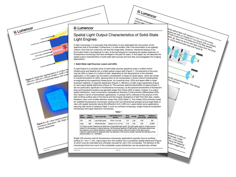
Thanks to its ability to access objects that are too small to be seen by the naked eye, microscopy has aided many discoveries and advancements across science and medicine. Not only have microscopes helped with disease discovery, they have also been used to identify key chemical compounds for drugs, and in today’s fast paced scientific landscape, they are becoming more important than ever. Microscopy’s ability to accurately capture the smallest details of biological samples is widely adopted in fields including life sciences, biotechnology, and pharmaceutical research.
Selecting the right light source technology is incredibly important, because it directly impacts the quality, precision, and versatility of imaging. Andrii Repula, Application Scientist at Lumencor explains: “The light source sits somewhat silently, like an unsung hero, but it’s crucial for the entire imaging ensemble.” Life sciences and material science in particular need accurate and uniform lighting in order to produce clear, detailed images of biological samples or materials used for semiconductor device fabrication.
Aspects such as the spectral, temporal, and spatial distribution of light can affect how researchers can visualise cells, tissues, or other microscopic structures. Spectral resolution refers to the wavelength of the light emitted by a source - different microscopy applications may require specific colours or wavelengths to achieve optimal results, and advanced light sources provide the flexibility to select and fine-tune these parameters. Temporal resolution, meanwhile, is about how light behaves over time. Many modern imaging systems are highly dynamic, and the light source must keep pace with changing requirements.
Spatial resolution is potentially the most critical when it comes to signal uniformity. This is about how light is distributed across the field of view, and can have a direct impact on the rendition of the image. Says Repula: “The spatial distribution of light should cover the entire sensor of the camera without uneven patches.”
The challenge: uniform illumination for high-quality imaging
One common issue faced by researchers and imaging professionals when it comes to spatial distribution, is non-uniform illumination across the field of view. Says Repula: “Anyone who wants to image cells or any biological sample wants to have the largest possible field of view with the most uniform illumination.”
Uneven lighting could degrade the quality of an optical image, particularly in the corners, where intensity often decays. To address this issue, the optical components, specifically the light sources, need to be able to ensure even light distribution, providing clearer, more reliable images for scientific analysis.
Addressing spatial resolution in microscopy
While spectral and temporal resolution have long been areas of attention in the development of light sources, spatial resolution has been gaining increasing attention. Repula pointed out that there has not historically been a wealth of shared data about the spatial distribution of light sources. “There are many light manufacturers out there, but I don't think there is much explicit data about spatial distribution,” he says.
A shortage of information can make it potentially difficult for researchers and industry professionals to select the right light source for their particular microscopy setup. The importance of understanding the numerical aperture - the measure of a system’s ability to gather light and resolve fine details at a given magnification - of both the light source and the camera sensor with which it is paired is particularly important.
Enter Lumencor, which provides this information for the benefit of its customers. Says Repula: “We explicitly show the numerical aperture of our solid light engines, whether they are LED, light pipes, or laser-based. This helps customers make informed decisions about which light source is the best fit for their camera and microscope setup.”
Matching light sources to imaging needs
One of the most significant advancements in light source technology is the ability to tailor the light to the specific requirements of the microscopy setup. Repula offers some insight into Lumencor’s range of solid light engines for microscopy, and how they are designed with different geometries and outputs to meet various imaging needs. “Lumencor manufactures products with either a liquid light guide or optical fibre,” he says. “We have light guides of different sizes - 3mm, 5mm - and different geometries of optical fibres like SMA (circular output) and FC/PC (square output). Lumencor is highly accomplished at designing other sizes and geometries to meet beam-shaping needs for our customers.”
By offering multiple options, researchers can ensure that the light source they select is best suited to their microscope and camera, whether they are working with small or large samples.
The ability to select and control colour is another important feature of advanced light sources. “Our light engines are colour-selective,” Repula explains. This means that researchers can easily choose the right colour of light to match their specific needs without having to rely on external filters. For example, violet light might be used to image cell nuclei, while yellow light is used to capture mitochondria.
Importantly, the spatial distribution remains consistent across different colours. “When customers take pictures with violet light and yellow light of the same field of view, the light distribution will be equally uniform,” says Repula. This consistency is particularly important for experiments involving multiple fluorescent dyes, as it ensures that the images of different dyes will align perfectly.
LED, light pipes, and lasers: tailored solutions for different applications
The unique characteristics of LEDs, light pipes, and lasers each offer different benefits depending on the application and the needs of the researcher. LED-based light sources, for instance, are ideal for imaging large samples where high resolution is not a highest priority. “LEDs and fluorescent light pipes have large angular distributions, making them great for imaging larger fields of view,” says Repula.
On the other hand, laser-based sources, with their more focused beams, are better suited for applications requiring fine detail. He continues: “Lasers have a smaller angular distribution and higher irradiance, making them perfect for super-resolution microscopy or when resolving fine details below the diffraction limit. Our goal is to offer light sources that fit seamlessly into most microscopy setups.” But that’s not all, Lumencor also offers custom solutions to ensure the highest level of compatibility.
Customisation: meeting researchers’ bespoke requirements
This customisation of Lumencor’s light engines and collimators to control the light distribution means, says Repula, that researchers do not need to compromise. Whether working on some sophisticated two-photon imaging or more conventional wide-field microscopy, scientists can more easily tailor their light sources to their unique applications.
He speaks from personal experience, following his days as a researcher trying to build optical setups from scratch. “Lumencor offers easy control of light,” he says. “With our compact light engines, researchers don’t need to build large, complex setups. Everything is combined into a small, user-friendly box.”
Whether imaging cells in a research lab or conducting drug studies in a pharmaceutical company, the right light source can make all the difference. Repula concludes by reiterating the importance of this often unsung hero: “After Lumencor, I have maybe one more favourite light source manufacturer, which is the universe itself. And our star, the Sun, is probably the most important light source for all of us. Looking at planet Earth, we can see how this light distribution that the Sun provides is important. When we have a lot of light in our sample, like at the Equator, we have one environment. When we have less light, like on the poles, we have totally different environmental conditions. This is why it is important to characterise the light distribution in any light source. At Lumencor, we do our best to develop perfect light sources that enable researchers to use light microscopy to push forward the frontiers of cell biology and neuroscience and translate those advances into diagnostics, therapeutics and clinical innovations.”


