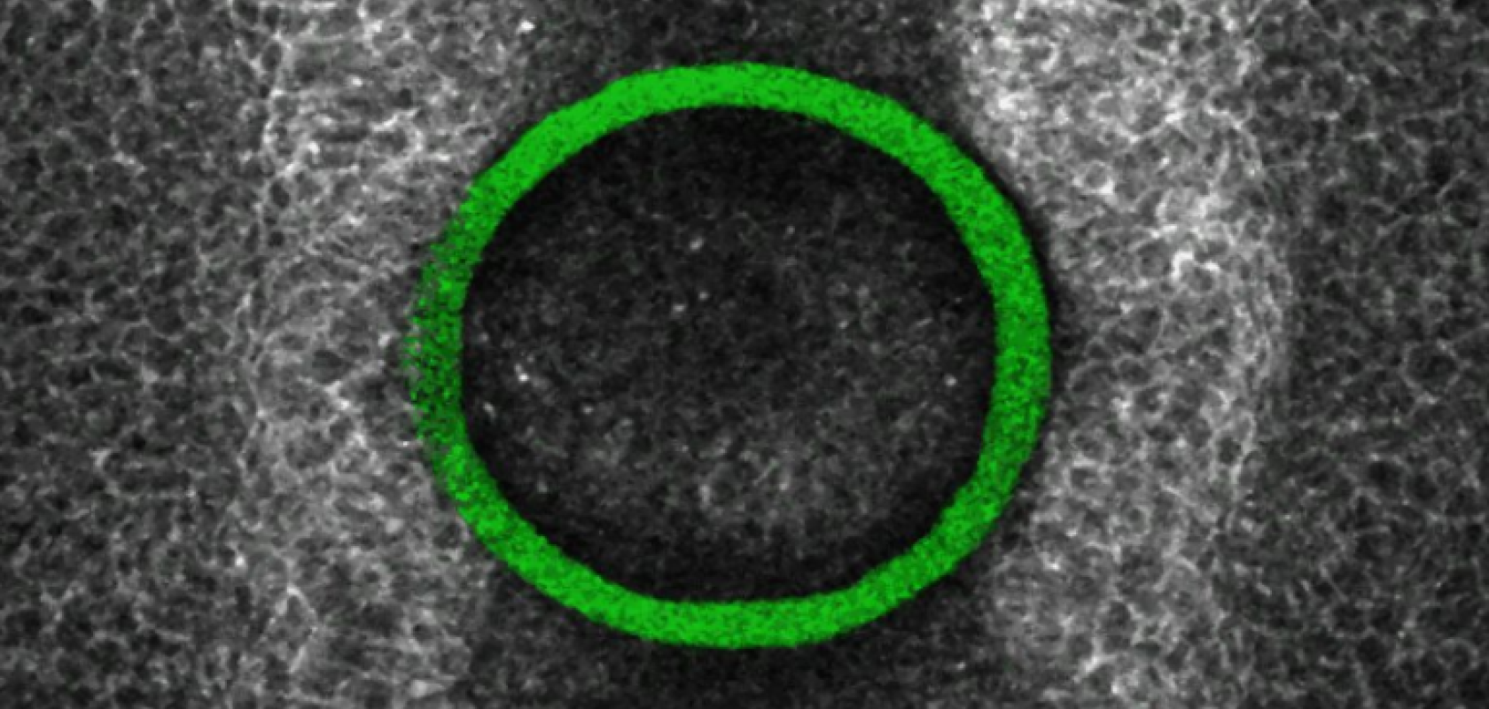
By positioning laser-printed mechanical force sensors directly in the developing brains and spinal cords of chicken embryos, scientists at University College London hope to understand and prevent birth malformations such as spina bifida.
In a study recently published in Nature Materials, research scientists from University College London (UCL), in collaboration with others at the University of Padua and the Veneto Institute of Molecular Medicine (VIMM), have used biotechnology sensors to measure the precise mechanical forces exerted on chicken embryos during development.
These forces are crucially important when organs and anatomical systems such as the neural tube, which creates the central nervous system, form. With congenital spinal cord malfunctions affecting one in 2,000 newborns in Europe alone each year, understanding them could be hugely impactful on medical science.
Understanding antenatal malformations
Although these types of malformations have been studied for decades, existing molecular and genetic research has been unable to fully understand them. Now, researchers are turning to the in-tissue mechanical forces that are present during embryo development. The challenge, however, is that as the embryonic spinal cord is so small – too small to be seen by the naked eye – and extremely delicate, any measuring device used to track development must be softened and miniaturised to such an extent as to avoid disrupting growth.
A small-scale solution to measure small-scale forces
To overcome this, researchers applied tiny 3D-printed liquid force sensors, about 0.1mm thick, directly inside developing chicken embryos. When the liquid sensor is exposed to a strong laser, it is then transformed into a spring-like solid, which attaches itself to the embryos’ growing spinal cord. The now-solid sensor then gets deformed by the mechanical forces produced by the embryo’s cells.
For embryonic development to occur and correctly form the spinal cord, these minute forces – roughly a tenth of the weight of a human eyelash – need to be greater than the opposing negative force. So the sensors’ ability to quantify forces inside the embryo would allow medical researchers to develop drugs that either increase the positive forces or decrease the negative ones sufficiently, and help prevent congenital malformations such as spina bifida.
“This technology is very versatile and widely applicable to many research fields,” said Dr. Gabriel Galea from UCL’s Great Ormond Street Institute of Child Health, who was a co-senior author of the study, “we hope it will be quickly adopted and applied by other groups to address fundamental questions.”
Another co-senior author and professor at the University of Padua and VIMM, Nicola Elvassore, said: “This discovery not only allows us to better understand the mechanical forces at play during embryonic development but also offers new perspectives for intervening and preventing conditions like spina bifada. The ability to quantify embryonic forces with such precision represents a significant step forward in biomedical research.”


